A couple week’s back I briefly described a complex anterior/posterior reconstruction we did for a very nice 54 yo woman from Florida (near Tampa).
This young lady had a Harrington Rod instrumentation and fusion done many years ago, and over the past 10+ years has developed progressive severe back pain, and trouble with increasing kyphotic posture.
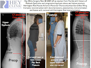
When you look at her preoperative clinical photograph, you can see that she stands with a typical “flat back” stance, with knees slightly flexed, but still is bent forward when you compare her torso to doorframe behind her. In addition, she has significant kyphosis above her previous fusion in thoracic spine area which is quite evident in preoperative photo, and X-Rays. In short, she has collapsed into kyphosis both above, and below her previous Harrington Rod fusion, and now also has spinal stenosis below her old fusion. Due to the severe progressive pain, she was on a significant amount of oral narcotic medication, which was becoming less and less effective.
To fix these problems, I started off anterior, removing L45 and L5S1 discs, and “jacking up” the anterior disc spaces with ALIF titanium cages and bone graft.
I then turned her over, after closing the anterior incision, and removed the Harrington rods.
I then did multiple posterior osteotomies in the thoracic and lumbar area, to allow for better correction of deformity, and then instrumented her spine from T3 all the way down to the sacrum and ilium with titanium alloy pedicle screw instrumentation and bone graft.
Her total surgical time was a little over 8 hrs for both the anterior and posterior combined procedures.
Postoperatively, she did very well, with both her and her family noticing her improved posture right away. She is also quite a bit taller, and can walk better due to better balance.
She went home to Florida about a week after surgery, and is doing well at home.
Her pre and postop X-Rays shown above show the marked imrovement in her “sagital balance”, or in layman’s terms, her balance from the side view. She is no longer pitched forward with flatback, and thoracic kyphosis. Her thoracic kyphosis is now normal, and her overall body shape is markedly improved. One way we measure the degree of sagital balance is by dropping a “plumb line” vertically from C7, and seeing where this line ends up relative to the L5S1 disc space. In a balanced person, C7 should sit over the top of the L5S1 disc space. I represent that plumb line with the red vertical liine. On the preoperative Xray, C7 sits over 12 cm in front of the L5S1 disc space. Postoperatively, the C7 sits normally over L5S1 disc.
When people are out of balance anteriorly, with “flat back syndrome” or significant kyphosis, it is very tiring and often painful. The pain, in part, is due to extreme muscle fatigue as the paraspinal and other muscles struggle to hold up the “Leaning Tower of Pisa” of the tilted torso. Many people find it hard to walk more than very short distances, since they often have to walk with their knees very slightly bent, which takes a huge amount of energy. Kyphosis and “Flat Back Syndrome” also greatly affects self-image and appearance in several ways: 1) Forward-tilting posture, 2) loss of normal lower lumbar/buttock contour (“no buttock syndrome”), and 3) pronounced thoracic “hump” in patients like this woman who have thoracic as well as lumbar kyphosis.
Lloyd A. Hey, MD MS
Hey Clinic for Scoliosis and Spine Surgery
Raleigh, NC USA
http://www.heyclinic.com


